Sonja's Dental History
"Sonja’s dental condition was serious, with many decayed, broken and infected teeth. While many cases are challenging, I would hope that no dentist ever again thinks that a 12 year-old child’s dental condition is hopeless." Dr. Lynn Mouden DDS, MPH
The images below are from Sonja Lauren’s past dental records.
Comments and descriptions have been provided by:
John E. Ward, DDS, MSD, FACP, Virginia Commonwealth University School of Dentistry
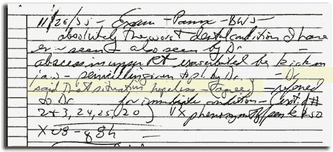
A portion of Sonja’s dental records from 11/26/75, before her teeth were extracted. Highlighted area reads: "Dr._____ said that situation hopeless — (agree)."
Radiograph of Sonja’s mouth before her natural teeth were extracted. The third molars (wisdom teeth) had not erupted into the mouth. The crowns of all the natural teeth are extensively affected by dental decay. However, the roots of the teeth and the bone level on the roots appear excellent. Some or all of the teeth could have been maintained by extensive and expensive dental procedures.

Radiograph
of Sonja’s mouth approximately 25 years after the natural teeth were
extracted. The bone which surrounded the natural teeth (alveolar bone)
has resorbed, which is a natural sequel to tooth loss, leaving the
basilar bone of the upper (maxilla) and lower (mandible) jaws. The
posterior ridge of the lower jaw has been increased in height by
surgically placing an artificial bone substitute (hydroxylapatite). Four
endosseous dental implants have been surgically placed in the bone at
the extension of the lower jaw. Attachments to supplement the retention
and stability of the lower complete denture project into the mouth from
two of the implants. The other two implants are submerged below the top
of the ridge.

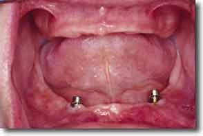
Sonja’s mouth illustrating healthy edentulous ridges and the attachments on the two implants.
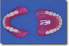
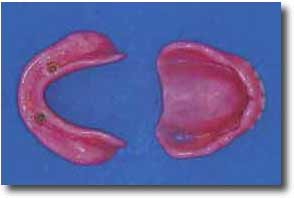
Three top (occlusal) view of Sonja’s upper and lower denture.
The
bottom (tissue surface) view of Sonja’s upper and lower denture. Note
the retentive devise of the two attachments imbedded in the denture
surface.
The upper and lower dentures in Sonja’s mouth.
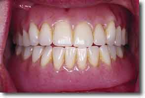
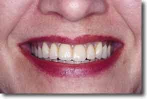
Sonja's beautiful smile!